Arachnoid Mater
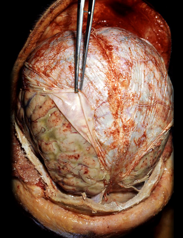
The arachnoid mater is a thin, transparent layer that surrounds the brain and spinal cord. It is connected to the pia mater by web-like filaments, hence the name “arachnoid” mater.
- Subarachnoid space. The space between the arachnoid mater and the pia mater in which CSF circulates. Many cerebral vessels course around the surface of the brain within the subarachnoid space.
- Arachnoid villi (granulations). Highly folded arachnoid mater that projects into the superior sagittal sinus and lateral lacunae (lateral extensions of the superior sagittal sinus). Arachnoid villi serve as sites where CSF diffuses into the superior sagittal sinus. Arachnoid villi often produce indentations in the inner surface of the calvarium.
 Generally, there is no space between the dura mater and the arachnoid mater. However, trauma to the head may stretch and rupture a bridging (cerebral) vein, resulting in bleeding into the subdural space (subdural hematoma). Because the damaged vessel is a vein, the increase in intracranial pressure and the effect of compressing the brain is much slower when compared to an epidural hematoma, which is caused by tearing of an artery. As a result, a subdural hematoma may develop over a period of days or even a week. Enlarging the subdural space is one factor that increases the risk of a subdural hematoma. As the subdural space enlarges, the bridging veins that traverse the space travel over a wider distance, causing them to be more vulnerable to tears. As a result, infants (who have smaller brains), the elderly (whose brains atrophy with age), and alcoholics (whose brains atrophy from alcohol use) are at increased risk of developing a subdural hematoma because of the tension of traversing vessels from the shrinking brain to the dural venous sinus. Subdural hematomas spread along the internal surface of the skull, creating a concave shape that follows the curve of the brain. The spread of blood is limited to one side of the brain due to dural reflections such as the tentorium cerebelli and falx cerebri. Contrast the spread of subdural hematomas to that of epidural hematomas that are limited in their spread due to the sutures.
A subarachnoid hemorrhage is defined as bleeding into the subarachnoid space because of a ruptured cerebral aneurysm.
Composed of dense collagenous connective tissue that is continuous with the dura mater of the spinal cord.
The dura mater envelopes the cranial nerves like a sleeve, which then fuse with the epineurium of the nerves outside the skull.
++++++++++++++++
-
in several areas the dura mater divides into two parts: a periosteal layer and a meningeal layer.
-
the meningeal layer of dura mater forms reflections that invaginate to support and protect various parts of the brain; the reflections also form the walls of the dural venous sinuses.
-
Falx cerebri
-
Tentorium cerebelli (Figure 2.3)
-
-
The two layers of dura mater are bound together. However, the layers separate at numerous locations to form dural septae or dural venous sinuses.
++Dural Septae
++Much like a seat belt assists in protecting a passenger from hitting the inside of the vehicle during an accident, four dural septae restrict displacement of the brain during everyday movements (Figure 15-4B).
- Falx cerebri. A sickle-shaped layer of dura mater between the cerebral hemispheres. The falx cerebri is attached anteriorly to the crista galli and posteriorly to the tentorium cerebelli. The superior and inferior sagittal sinuses form the superior and inferior margins. The inferior edge of the falx cerebri courses along the superior surface of the corpus callosum.
- Tentorium cerebelli. Separates the occipital lobes of the cerebrum from the cerebellum. The tentorium cerebelli encloses the transverse sinuses posteriorly and the superior petrosal sinuses anteriorly (Figure 15-4C).
- Falx cerebelli. A small triangular extension of the tentorium cerebelli containing the occipital sinus. The falx cerebelli descends, separating the cerebellar lobes, and terminates at the foramen magnum.
- Diaphragma sellae. A circular horizontal fold of dura that covers the pituitary by forming a roof over the sella turcica.
The dural sinuses are grouped into the sagittal, lateral (including the transverse, sigmoid, and petrosal sinuses), and cavernous sinusesThe dural venous sinuses are venous channels located between the periosteal and the meningeal layers of the dura mater. The dural venous sinuses are lined with endothelium and lack valves. They serve as a receptacle for blood from the cerebral, diploic, and emissary veins. They also receive the cerebrospinal fluid (CSF), drained by the arachnoid granulations. Blood in the dural venous sinuses primarily drains into the internal jugular veins.
- Superior and inferior sagittal sinuses. Course in the midline along the superior and inferior borders of the falx cerebri. The inferior sagittal sinus joins with the great cerebral vein (of Galen) to form the straight sinus.
- Straight sinus. Courses along the line of attachment of the falx cerebri to the tentorium cerebelli.
- Occipital sinus. Courses within the falx cerebelli.
- Confluence of sinuses. Forms the union of the superior sagittal straight and occipital sinuses and is located deep to the internal occipital protuberance.
- Transverse sinus. Courses laterally from the confluence of sinuses within the tentorium cerebelli.
- Sigmoid sinus. A continuation of the transverse sinus and descends in an S-shaped groove to join with the inferior petrosal sinus at the jugular foramen, forming the internal jugular vein.
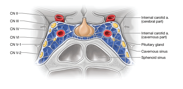
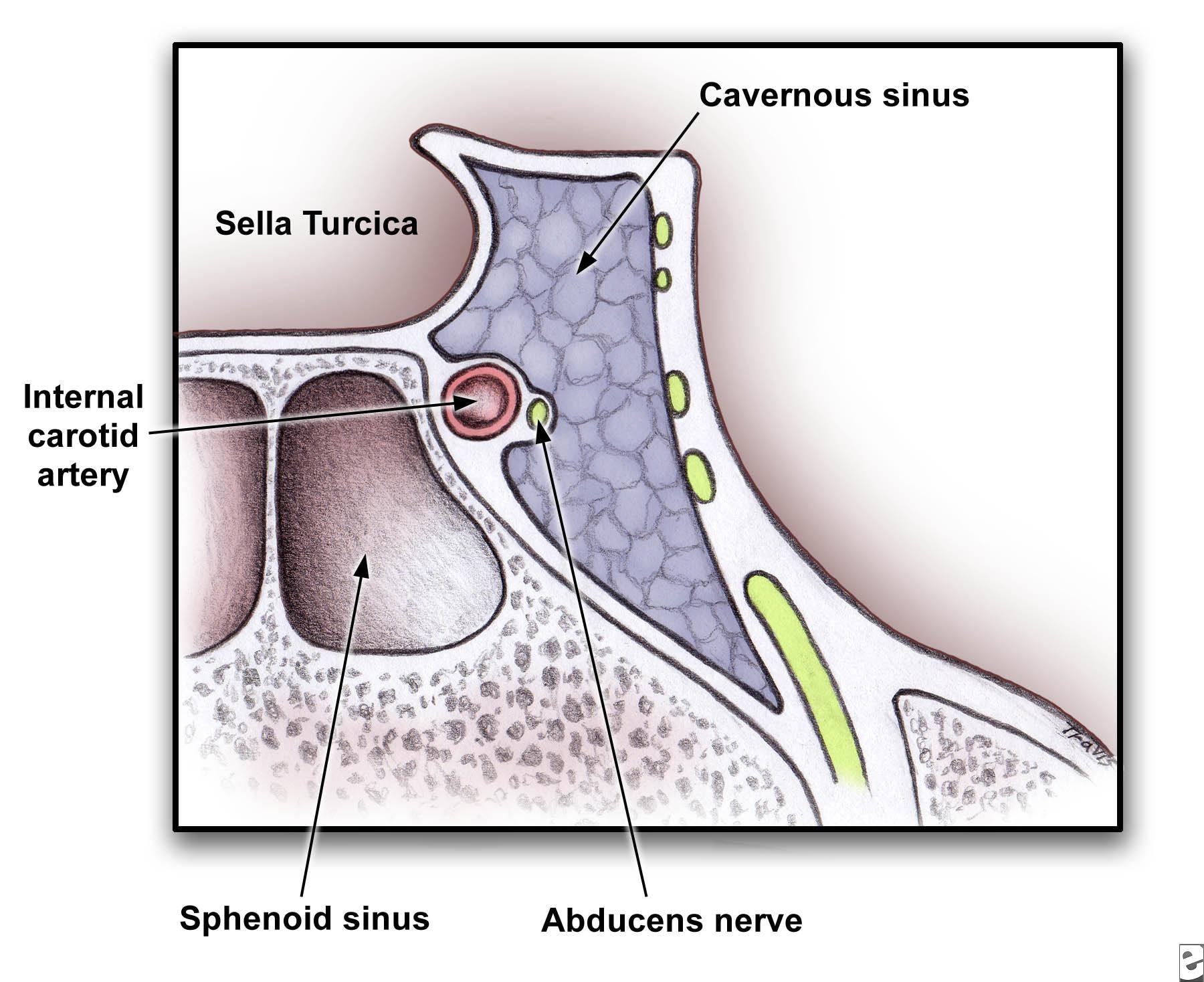
- Cavernous sinus. Located on each side of the sella turcica. The internal carotid artery and CN VI course through the middle of the sinus. Cranial nerves (CNN) III, IV, V-1, and V-2 course anteriorly through the lateral walls of the sinus. The cavernous sinus communicates with the pterygoid venous plexus via emissary veins and the superior and inferior ophthalmic veins.
 Anatomy of cross section of cavernous sinus showing close proximity to cranial nerves and sphenoid sinus.
The cavernous sinus is the only location in the body where an artery courses through a venous structure. A carotid-cavernous sinus fistula forms when the internal carotid artery ruptures within the cavernous sinus.
++Pituitary tumors can expand in the direction of least resistance and compress the cavernous sinus structures (CNN III, IV, V-1, V-2 and VI), causing paralysis of the extraocular muscles and sensory loss in the forehead and maxillary region.
- Superior petrosal sinus. Courses along the superior portion of the petrous part of the temporal bone and drains into the transverse sinus.
- Inferior petrosal sinus. Courses along the inferior portion of the petrous part of the temporal bone and drains into the cavernous sinus. The inferior petrosal and sigmoid sinuses unite to become the internal jugular vein.
- Diploic veins. Course within the spongy portion of cranial bones.
- Emissary veins. Course between the scalp and dural venous sinuses.
-
++
Pia Mater
The pia mater is the most internal and delicate of the meninges surrounding the brain and spinal cord (Figure 15-4D). The pia mater forms a sheath around blood vessels as they course into the fissures and sulci and penetrate the brain. The pia mater joins with the ependymal cells that line the ventricles of the brain to form choroid plexuses that produce CSF.